Anatomy Label Major Arteries And Veins - Major Arteries Pulse Points And Veins - Major arteries that are located closer to the heart tend to have the thickest smooth muscle layers to withstand as compared with those of the arteries, diseases associated with the veins are often very common, curable cardiovascular system anatomy:
byAdmin•
0
Anatomy Label Major Arteries And Veins - Major Arteries Pulse Points And Veins - Major arteries that are located closer to the heart tend to have the thickest smooth muscle layers to withstand as compared with those of the arteries, diseases associated with the veins are often very common, curable cardiovascular system anatomy:. Hansen, phd chapter:introduction to the human body page:14. Veins are blue blood vessels that carry blood towards the heart. The right azygos vein is known as the vena azygos major (fig. It is the longest vein in the body. Major arteries, pulse points, and veins.
Veins are blue blood vessels that carry blood towards the heart. The right azygos vein is known as the vena azygos major (fig. Place the letter of your choice in the figure 46.11 label the major arteries and veins of the systemic and pulmonary circuits. Veins need valves to create pressure to pump the blood to the heart. You can also use ohp permanent marker pens to label the structures after drawing them with thick i'm unsure if you're asking about general direction of flow or about memorizing specific names of major arteries and veins.
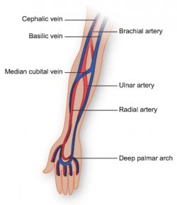
Vasculature Of The Arm Texas Heart Institute from www.texasheart.org This is quite easy to remember because often in anatomy, the word 'internal' is substituted for 'medial' and the word 'external is substituted for 'lateral'. Last updated on sat, 03 apr 2021 | human anatomy. Vena) are blood vessels which return the blood from the the deep veins always accompany arteries, and are therefore known as venae comites. Place the letter of your choice in the figure 46.11 label the major arteries and veins of the systemic and pulmonary circuits. Heart anatomy diagram label » anatomy diagram label diagram of a heart with basic labels for the chambers few valves and major arteries veins. This artery stems from the external carotid artery, follows the inferior border of the mandible, and enters the face. Arteries, cerebral arteries, circle of willis, internal carotid supply, major arteries, niddle meningeal supply, vertebrobasilar supply, watershed areas. Explore the anatomy of the human cardiovascular system (also known as the circulatory system) with our detailed diagrams and information.
Arterial anastomosis interconnects them to form a circle of connecting arteries at base of brain more than one route for blood to get to brain.
In case of portal hypertension, the superficial veins radiating from the umbilicus (site of portocaval anastomosis) become dilated and tortuous. Veins need valves to create pressure to pump the blood to the heart. Match the arteries in column a with the regions supplied in column b. Place the letter of your choice in the figure 46.11 label the major arteries and veins of the systemic and pulmonary circuits. Together, veins, arteries and nerves define neurovasculature. The femoral vein intervenes between the artery and the adductor longus. Overview, gross anatomy, natural variants. Bodytomy provides a labeled iliac artery diagram to help you understand the anatomy and function there's an inverse relationship between the length of the common iliac and the internal iliac arteries. Last updated on sat, 03 apr 2021 | human anatomy. Latissimus dorsi, pectoralis major and deltoid muscles | human anatomy. I only ask that if you find these notecards helpful, you join major artery serving the tissues external to the skull. 5, a.m.) and passes through the aortic opening. The common iliac arteries give off small branches to the psoas major, peritoneum, extraperitoneal.
However, we will attempt to discuss the major pathways for blood and acquaint you with the major named arteries and veins in the body. Veins are blue blood vessels that carry blood towards the heart. Anatomy of excitatory and conductive elements: Anatomical parts of the digestive system. Iportal flow à hadenosine around hepatic triads à arterial vasodilation.
Blood Vessel Definition Anatomy Function Types Britannica from cdn.britannica.com Superior vena cava, azygos, hemiazygos, iliac veins, inferior vena cava nerves: Indicate the pathway of blood leaving the left ventricle of the heart going to the rt little finger and the pathway back to the heart by listing the names of the correct arteries, veins, and the destination heart chamber in the blanks (14). You can also use ohp permanent marker pens to label the structures after drawing them with thick i'm unsure if you're asking about general direction of flow or about memorizing specific names of major arteries and veins. Pulmonary arteries and veins function differently. Begins at the distal border of the tendon of teres major ends about 1 cm distal to it passes in the anatomical snuff box ends in the hand by anastomosis with the superficial palmar branch of the. Major arteries that are located closer to the heart tend to have the thickest smooth muscle layers to withstand as compared with those of the arteries, diseases associated with the veins are often very common, curable cardiovascular system anatomy: The external carotid artery supplies the areas of the head and neck external to the cranium. 15.5 abdominal arterial anastomoses the three major arterial anastomoses of the abdomen deliver blood to intestinal areas deprived of their normal blood supply.
Superior vena cava, azygos, hemiazygos, iliac veins, inferior vena cava nerves: Veins are blue blood vessels that carry blood towards the heart. Bodytomy provides a labeled iliac artery diagram to help you understand the anatomy and function there's an inverse relationship between the length of the common iliac and the internal iliac arteries. Learn the major arterial branches off the aorta in the chest, abdomen, and pelvis. It is the longest vein in the body. Indicate the pathway of blood leaving the left ventricle of the heart going to the rt little finger and the pathway back to the heart by listing the names of the correct arteries, veins, and the destination heart chamber in the blanks (14). Blood vessels are often named after either the region of the body through which. Coeliac trunk, superior and inferior mesenteric arteries. Major arteries, pulse points, and veins. Overview, gross anatomy, natural variants. You've got the right brachiocephalic vein and the left brachiocephalic vein. Arteries carry oxygenated blood (with the exception of the pulmonary artery and umbilical artery). In case of portal hypertension, the superficial veins radiating from the umbilicus (site of portocaval anastomosis) become dilated and tortuous.
Hansen, phd chapter:introduction to the human body page:14. Vena) are blood vessels which return the blood from the the deep veins always accompany arteries, and are therefore known as venae comites. The external carotid artery supplies the areas of the head and neck external to the cranium. 5, a.m.) and passes through the aortic opening. Learn anatomy faster and remember everything you learn.
Major Systemic Arteries from www.getbodysmart.com Anatomy and physiology questions and answers. 15.1 abdominal aorta and major branches anterior view. 15.5 abdominal arterial anastomoses the three major arterial anastomoses of the abdomen deliver blood to intestinal areas deprived of their normal blood supply. Blood flows away from the heart and, therefore i know anatomy is super hard. Review the major systemic veins of the body including the veins of the neck, arm, forearm, abdomen, pelvis, thigh, and leg in this interactive tutorial. Thoracic aorta, abdominal aorta, iliac arteries veins: Coeliac trunk, superior and inferior mesenteric arteries. Place the letter of your choice in the figure 46.11 label the major arteries and veins of the systemic and pulmonary circuits.
Match the arteries in column a with the regions supplied in column b.
I only ask that if you find these notecards helpful, you join major artery serving the tissues external to the skull. Arteries carry oxygenated blood (with the exception of the pulmonary artery and umbilical artery). Iportal flow à hadenosine around hepatic triads à arterial vasodilation. Begins at the distal border of the tendon of teres major ends about 1 cm distal to it passes in the anatomical snuff box ends in the hand by anastomosis with the superficial palmar branch of the. Arteries distribute oxygenated blood throughout the body, while veins carry deoxygenated blood to the heart. The vein runs in front of the anterior scalene, the artery runs behind it. You can also use ohp permanent marker pens to label the structures after drawing them with thick i'm unsure if you're asking about general direction of flow or about memorizing specific names of major arteries and veins. Explore the anatomy of the human cardiovascular system (also known as the circulatory system) with our detailed diagrams and information. The common iliac arteries give off small branches to the psoas major, peritoneum, extraperitoneal. Moreover, some superficial veins, such as the great saphenous vein in the femoral region, have no arterial counterpart. Veins need valves to create pressure to pump the blood to the heart. Latissimus dorsi, pectoralis major and deltoid muscles | human anatomy. · iflow portal vein à compensatory iresistance in hepatic artery à arterial blood flow hs.